Neurons
Neurons are cells that are responsible for electrical and chemical communication in the central nervous system through specialized connections known as synapses. The central nervous system is estimated to contain an astounding 80-100 billion neurons and can process millions of bits of information each minute to coordinate appropriate responses. This information is transmitted in the form of action potentials – or nerve impulses – through a succession of neurons. These impulses may, however, be blocked, changed, or integrated along the way at the synapses.
Formation and Structure
Neurons are embryologically derived from the central part of the ectoderm – the neural plate – by a process called neurogenesis. Neuroblasts differentiate into many types of cells by genetically predetermined migration. These neurons can be found in many different locations and have varying sizes and shapes that determine their function. They do not necessarily have a generic structure, but all neurons have a cell body, an axon, an axon terminal, and dendrites.
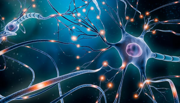
The circular cell body, or soma, varies in size from 4-100 µm, and contains the nucleus. This opens into an elongated stem-like structure, called an axon or synaptic terminal bulb, which ends in an axon terminal. The cell body narrows like a funnel at the junction between the soma and axon; this is called the axon hillock and most action potentials are generated here. There are extensions on the surface of the cell body called dendrites that are the main receptive organelles of the neuron. The number of these dendritic extensions determines how neurons are classified – whether they are multipolar, unipolar, or pseudounipolar.
Nerve degeneration and regeneration require active transport in and out of the cell, a function that is achieved by the microtubular proteins kinesin and dynein. Failure of these mechanisms or an imbalance between neuronal formation and loss has been associated with clinical states such as Parkinson’s. Another significant group of conditions that is linked to nerve cell damage is infections; viruses like polio, rabies, herpes simplex and varicella zoster infect nerve terminals, migrate up to the cell body into the nucleus by way of microtubular proteins, and multiply there destroying the neurons. Some viruses, notably varicella zoster, stay dormant in the cell body for a variable amount of time and reactivate during periods of immunosuppression to cause disease states like shingles.
Functions of Neurons
The neuron acts as a portal for both receiving and sending information, by way of a highly complex mechanism involving neurotransmitters and action potentials. The dendrites of a neuron generate excitatory and inhibitory post-synaptic potentials by depolarization and repolarization respectively, when stimulated by neurotransmitters, hormones, drugs, light, or other neurons. This information is then integrated, decoded, and sent as a chemical or electric signal via neuronal connections. Neurons can be classified according to their functionality as sensory or afferent, motor or efferent, and interneurons or relay neurons. The structure of neurons at specific sites in the brain and spinal cord vary according to their primary function.
Emerging Research
The neuronal protein α-synuclein is currently being investigated for its potential as a biomarker to predict and prognosticate Parkinson’s disease, for which there is currently no available alternative.1 In a similarly ground-breaking field, a recent study looking at the regeneration of peripheral nerves concluded that mesenchymal stem cells derived from Wharton’s jelly (WJ-MSCs) can be transformed into Schwann cell-like cells (WJ-SCLCs) that may play a key role in the development of therapeutic possibilities for disorders associated with nerve injury or loss.2
Research shows that certain specific neuronal cell proteins can be used as markers for brain injury. In particular, glial fibrillary acidic protein (GFAP) and ubiquitin c-terminal hydrolase L1 (UCH-L1) have both been recently approved by the FDA for this application as they have a high sensitivity for detecting acute intracranial lesions on imaging.3
References
1. Fu Y, Jiang C, et al. (2020) Facile Impedimetric Analysis of Neuronal Exosome Markers in Parkinson's Disease Diagnostics. Anal Chem. 92(20):13647-13651. https://pubs.acs.org
2. Choi SJ, Park SY, et al. (2021) Mesenchymal Stem Cells Derived from Wharton's Jelly Can Differentiate into Schwann Cell-Like Cells and Promote Peripheral Nerve Regeneration in Acellular Nerve Grafts. Tissue Eng Regen Med. 18(3):467-478. https://link.springer.com
3. Bramblett GT, Harris JN, et al. (2021) Traumatic Optic Nerve Injury Elevates Plasma Biomarkers of Traumatic Brain Injury in a Porcine Model. Journal of Neurotrauma.38(8):1000-1005. https://doi.org/10.1089/neu.2020.7039
