Natural Killer Cells
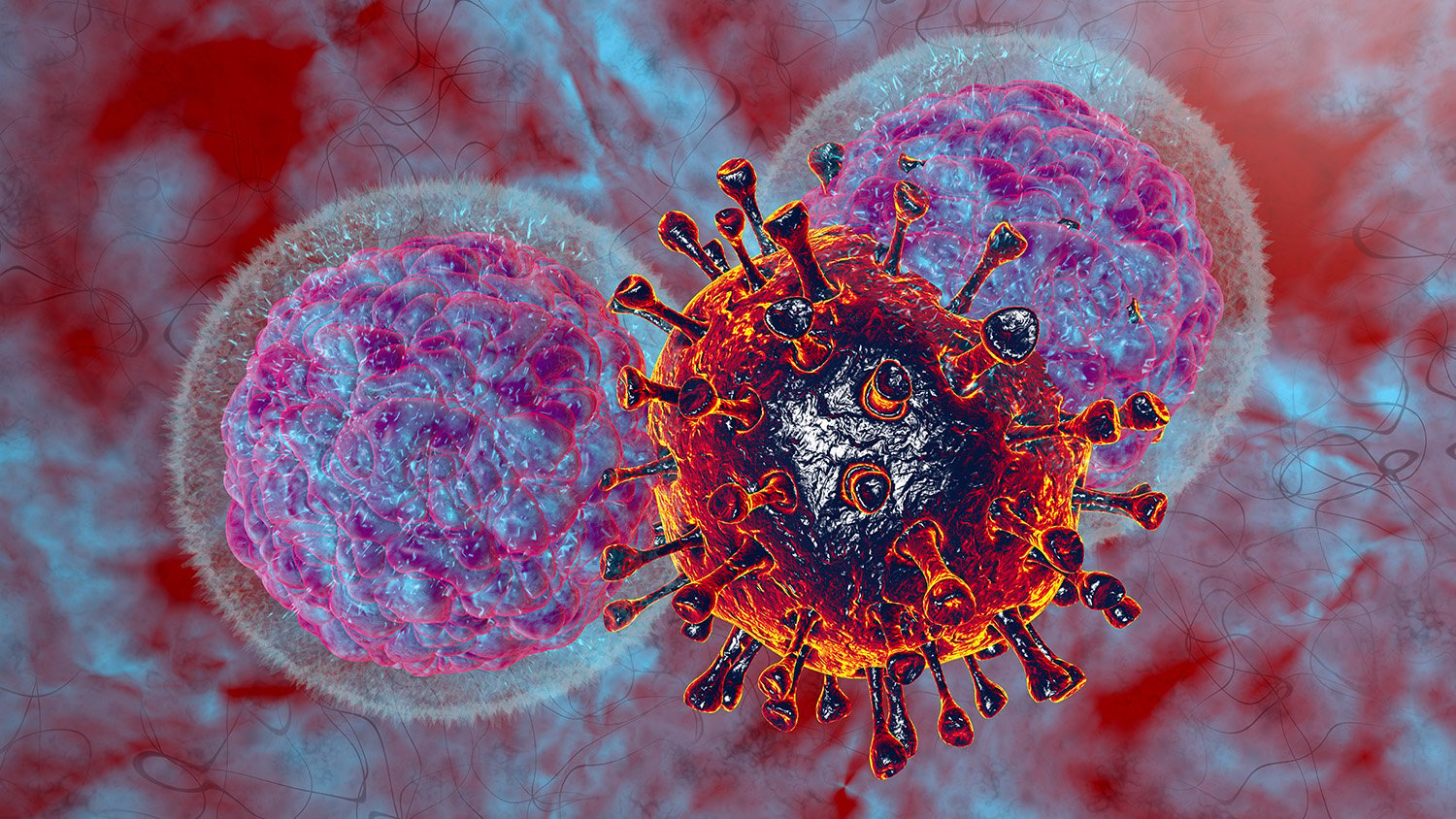
Natural killer (NK) cells are a type of innate lymphoid cells that lack antigen specificity but cause spontaneous lysis of physiologically stressed cells, such as tumor cells and virus-infected cells, without prior sensitization. Activated NK cells also modulate the functions of other innate and adaptive immune cells by secreting several cytokines such as interferon-γ (IFN-γ), tumour necrosis factor-α (TNF-α), granulocyte macrophage colony stimulating factor (GMCSF), and chemokines. They can recognize target cells by the absence of surface MHC class I molecules1, by stress-associated molecules (e.g. ULPB) or antibodies bound to the surface of the target cell (antibody-dependent cellular cytotoxicity, ADCC). The development of engineered NK cells for the treatment of cancer is gaining a lot of interest in clinical research.
Formation and structure
NK cells are formed from the common lymphoid progenitors (CLPs) in the bone marrow but can also be generated by secondary lymphoid tissues including the spleen, tonsils, and lymph nodes. CD34+ hematopoietic stem cells differentiate into lymphoid-primed multipotential progenitors (LMPP): they subsequently transition into CPLs by expressing CD38, CD7, CD10, and CD12, which have the potential to differentiate into many types of innate lymphoid cells. Expression of CD122 commits the CLPs into NK cell lineage and then CD56 marks the transition of an immature NK cell to a mature state.2
Under light microscopy, the morphology of NK cells is like that of a large granular lymphocyte, with big nuclei that contain coarse chromatin and prominent nucleoli. The cytoplasm of these cells stains basophilic with azurophilic granules. Electron microscopic (EM) examination suggests that NK cells exhibit various shapes and microvilli, and their granules show structural and functional similarities to lysosomes.3
Function
As the name suggests, NK cells are potent natural killers. Healthy cells express MHC class I molecules which induce an inhibitory signal in NK cells, and the absence or downregulation of MHC class I on tumor or virus-infected cells results in activation of NK cells. Also, the recognition of stress-induced ligands such as MHC class I polypeptide-related sequence A and B (MICA and MICB) - expressed on the surface of target cells – by NK cells triggers cytotoxic activity. NK cells lack the T cell receptor but can induce apoptosis of cells to which antibody molecules have attached through a process called antibody-dependent cellular cytotoxicity (ADCC) by binding to the Fc portion of the antibody.1, 4 Lysis of target cells is achieved by the following three mechanisms:
- NK cell activation produces INF-γ which in turn activates macrophages promoting opsonization.
- ADCC through the interaction of CD16 with cell-bound IgG
- direct killing via secretion of perforin and granzymes A, B, and K.1, 3
The signaling pathway for NK cell activation relies on a dynamic balance between the activating and the inhibiting receptors, which in context of the target HLA molecule profile, determines the vulnerability of the target cells to be attacked by a given NK cell. This has significant clinical relevance in the management of organ transplants and viral infections. A diverse range of receptors is expressed on NK cells, which are genetically predetermined by polymorphic genes present on chromosomes 12 and 19.1
Activating receptors:
- NKG2D/CD314 triggers NK cell activation by recognizing stress-induced ligands – MICA, MICB, and ULBP1-6 – expressed on damaged and abnormal cells and the secretion of IF-α and IF-β
- NKG2C/CD94 provides activating signals for NK cells by engaging with HLA-E expressed on target cells (please see NGG2A/CD94 under inhibitory receptors)
Inhibiting receptors:
- NKG2A/CD94 provides inhibitory signaling for NK cells by engaging with HLA-E expressed on target cells
- KIRs signal inhibitory pathway by binding with HLA- A, B, and C expressed on target cells
The function of NK cells is compromised in many acquired and congenital immune deficiency disorders. These patients are highly susceptible to viral infections, for example, human papillomavirus, herpes simplex, and cytomegalovirus.
Emerging research
While chimeric antigen receptor engineered T cells (CAR-T) have already been successfully used for the treatment of hematologic malignancies, the use of CAR-NK cells to fight cancer is still in early phases of investigation. Employing engineered NK cells has significant advantages over using T cells because these cells do not elicit graft versus host disease; hence these can be used in allogenic settings.5 The genomic editing with the CRISPR/Cas9 system is expected to fully potentiate NK cell immunotherapy in the future, by arming NK cells with CAR constructs, enhancing activating pathways, facilitating NK cell infiltration into tumours, and by antagonizing the inhibitory pathways.6
Studies to develop methods for activating genetically non-modified NK cells using cytokines to further enhance therapeutic efficacy are underway.7 Developing immunomodulatory drugs to use in combination with targeting antibodies can enhance the function of NK cells and can prove helpful in promoting ADCC responses for the treatment of cancer. In the future, strategies to exploit NK cells for vaccine development and management of autoimmune diseases – including recurrent pregnancy loss – are being explored.8, 9
Tools to study NK cells
NK cells can be identified by flow cytometric assessment of specific cell markers in peripheral blood, cord blood, and cell suspensions from placental tissues as well as other body compartments. Understanding the molecular mechanisms between activating and inhibitory signals is challenging, however, newer technologies such as ligand-functionalized nanodot arrays can help to translate the interactions between receptors. The ultrastructure of activated cells can be analyzed by transmission electron microscopy, and the newer microfluidics technologies can help to understand NK cell heterogeneity.10, 11
Single-cell analysis tools help to understand the vast diversity of NK cell phenotypes and high-resolution imaging allows researchers to analyze the spatial context of NK cells in combination with other methods, such as single-cell sequencing and spatial transcriptomics. Many diverse subsets of NK cells can be identified by single-cell RNA sequencing (scRNA-seq) encompassing both known and novel types.12
Cell markers
NK cells are typically identified by the presence of CD56 and the absence of CD3 and TCR. More mature and potentially cytotoxic NK cells are CD56-/CD16+ and less mature cells are CD56+/CD16-.1 The evaluation of the following markers is crucial for studying NK cells.10, 12
| Surface marker | Alternative names | Location |
|---|---|---|
| CD16a | FcyRIIIA, FCGR3A, FCG3, FCGR3, IGFR3 | Surface |
| CD25 | IL-2RB | Surface |
| CD56 | neural cell adhesion molecule 1 | Surface |
| CD62L | L-Selectin | Surface |
| CD335 | NCR1, NK-p46 | Surface |
| CD336 | NCR2, NK-p44 | Surface |
| CD337 | NCR3, NK-p30 | Surface |
| NKp80 | Clec5C | Surface |
| CD314 | NKG2D, KLRK1 | Surface |
| CD352 | NTB-A, SLAMF6 | Surface |
| CD253 | Apo-2L | Surface |
| CD247 | TCR, Z Chain | Surface |
| CD244 | NAIL, 2B4 | Surface |
| CD223 | LAG-3 | Surface |
| CD18b | IL18Rβ, IL18RAP | Surface |
| CD218a | IL18R1 | Surface |
| CD158a | KIR2DL1 | Surface |
| CD158b1 | KIR2DL2 | Surface |
| CD158b2 | KIR2DL3 | Surface |
| CD158c | KIR3DP1 | Surface |
| CD158d | KIR2DL4, KIR-103AS | Surface |
| CD158e | KIR3DL1 | Surface |
| CD158f1 | KIR2DL5A | Surface |
| CD158f2 | KIR2DL5B | Surface |
| CD158g | KIR2DS5 | Surface |
| CD158h | KIR2DS1 | Surface |
| CD158i | KIR2DS4 | Surface |
| CD158j | KIR2DS2 | Surface |
| CD158k | KIRDL2 | Surface |
| CD158z | KIR3DL3 | Surface |
| CD159a | NKG2A, KLRC1 | Surface |
| CD159c | NKG2C, KLRC2 | Surface |
| CD160 | BY55 | Surface |
| CD161 | NKR-P1A, KLRB1 | Surface |
| CD122 | IL2RB | Surface |
| CD94 | KLRD1, KP43 | Surface |
| CD328 | Siglec-7, AIRM-1 | Surface |
| TIGIT | VSTM3 | Surface |
| CD137 | 4-1BB | Surface |
| CD279 | PD-1 | Surface |
| CD274 | PD-L1 | Surface |
References
1. Sourav Paul, Girdhari Lal. (2017). The molecular mechanisms of natural killer cells function and its importance in cancer immunotherapy. Front. Immunol. 8:1124. https://doi.org/10.3389/fimmu.2017.01124
2. Abel Alex M., Yang Chao et al. (2018) Natural Killer Cells: Development, Maturation, and Clinical Utilization. Frontiers in Immunology. 9:1869. doi:10.3389/fimmu.2018.01869
3. Rahman M, Bordoni B. (2021) Histology, Natural Killer Cells. StatPearls Publishing. https://www.ncbi.nlm.nih.gov/books/NBK565844/
4. Activating Macrophages and NK Cells. (2021). Community College of Baltimore Country (Cantonsville). https://bio.libretexts.org/@go/page/3329
5. Xie Guozhu Han Dong et al. (2020) CAR-NK cells: A promising cellular immunotherapy for cancer. EBioMedicine. 59:102975. doi:https://doi.org/10.1016/j.ebiom.2020.102975
6. Afolabi, L.O., Adeshakin A.O. et al. (2019). Genetic reprogramming for NK cell cancer immunotherapy with CRISPR/Cas9. Immunology. 158:2. doi.org/10.1111/imm.13094
7. Xu, J., Niu, T. Natural killer cell-based immunotherapy for acute myeloid leukemia. J Hematol Oncol 13, 167 (2020). https://doi.org/10.1186/s13045-020-00996-x
8. Cox A, Cevik H, Feldman HA et al. (2021). Targeting natural killer cells to enhance vaccine responses. Trends Pharmacol Sci. 42(9):789-801. doi: 10.1016/j.tips.2021.06.004. PMID: 34311992; PMCID: PMC8364504.
9. Kucuksezer UC, Aktas Cetin E et al. (2021). The Role of Natural Killer Cells in Autoimmune Diseases. Front Immunol. 12:622306. doi: 10.3389/fimmu.2021.622306. PMID: 33717125; PMCID: PMC7947192.
10. Liu, S., Galat, V. et al. (2021). NK cell-based cancer immunotherapy: from basic biology to clinical development. J Hematol Oncol. 14:7. https://doi.org/10.1186/s13045-020-01014-w
11. Le Saux, G., & Schvartzman, M. (2019). Advanced Materials and Devices for the Regulation and Study of NK Cells. International journal of molecular sciences, 20(3):646. doi.org/10.3390/ijms20030646
12. Samantha L. Smith, Philippa R. et al. (2020). Diversity of peripheral blood human NK cells identified by single-cell RNA sequencing. Blood Adv. 4 (7): 1388–1406. doi: https://doi.org/10.1182/bloodadvances.2019000699
