Precision performance of the DxFLEX ClearLLab 10C application system
Sample Preparation

Integrated FDA cleared and CE-marked IVD leukemia and lymphoma* (L&L) immunophenotyping solution for lymphoid and myeloid lineages
*For Non-Hodgkin’s lymphoma only
Sample Acquisition
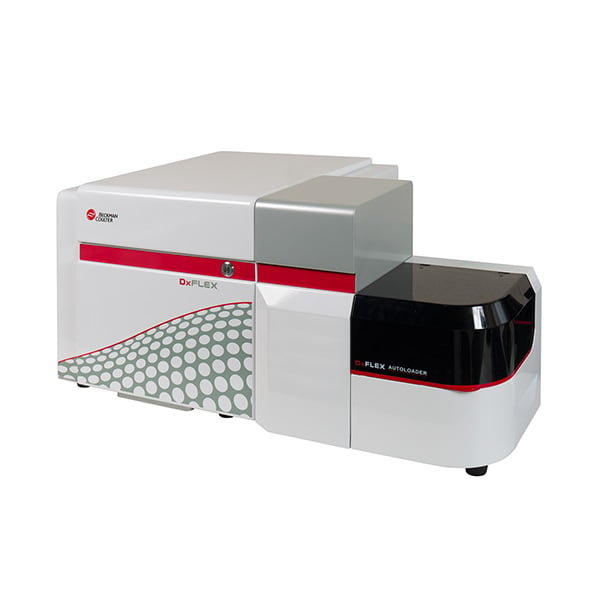
The DxFLEX Flow Cytometer streamlines high-complexity testing with a 13-color** capability and new detector technology for easier compensation.
**In the US, the DxFLEX flow cytometer is cleared for 10-color in vitro diagnostic use with the ClearLLab 10C Reagent System. Fluorescence channels FL11-FL13 and all other applications are for Research Use Only.
Sample Analysis

Flow Cytometry Analysis Software
Purpose-built for clinical laboratories, Kaluza C Flow Cytometry Analysis Software helps you turn critical data into meaningful patient reports.
Introduction
Flow cytometry immunophenotyping (FCI) is a valuable diagnostics tool for hematolymphoid malignancies.1
The DxFLEX* flow cytometer is the clinical version of the CytoFLEX flow cytometer. Derived from the successful research platform, the DxFLEX flow cytometer has the advantage of using Avalanche Photodiode (APD) technology, which has higher sensitivity than typical Photomultiplier Tube (PMT)-based cytometers and makes high-color flow cytometry testing more accessible because there is a linear correlation between gain setting/voltages and measured fluorescence intensities.2, 3
By leveraging the ClearLLab 10C** application, a multisite clinical study was conducted by using DxFLEX flow cytometers to demonstrate the precision performance of the DxFLEX ClearLLab 10C as a complete system.
* In the US, the DxFLEX flow cytometer is cleared for 10-color in vitro diagnostic use with the ClearLLab 10C Reagent System. Fluorescence channels FL11-FL13 and all other applications are for Research Use Only.
** The ClearLLab 10C Reagent System on the DxFLEX flow cytometer has received 510k clearance and IVDR certification.
Results
Precision using ClearLLab Control Cells
Targeted populations as those defined in the assay sheet for ClearLLab Control Cells were evaluated for precision performance on the DxFLEX ClearLLab10C system. Both ClearLLab Control Cells, Normal and Abnormal, were tested using the ClearLLab 10C B, T, M1 and M2 panels. A total of 140 replicates per each panel for each level of control cells were collected and analyzed using Kaluza C software. The overall mean percent recovery of the targeted populations in the B panel ranged from 7.99% to 97.92%, in the T panel ranged from 2.77% to 97.83%, in the M1 panel ranged from 7.70% to 97.88%, and in the M2 panel ranged from 7.96% to 98.03%. For each targeted population, the overall repeatability from both levels ranged from 0.24% CV to 5.56% CV, and the reproducibility ranged from 1.00% CV to 16.93% CV. The representative results from using the Abnormal Control Cells are shown in Table 1 for B panel, Table 2 for T panel, Table 3 for M1 panel, and Table 4 for M2 panel. Although data is not shown, Normal ClearLLab Control Cells demonstrated similar performance. In addition, each individual site had satisfying precision performance on each individual DxFLEX instrument, with repeatability from both levels ranging from 0.01 % CV to 9.49 % CV and variation within instrument ranging from 0.02% CV to 12.16% CV.
| Name | N | Mean | Repeatability | Between runs | Between days | Between instruments | Reproducibility | |||||
| SD | CV% | SD | CV% | SD | CV% | SD | CV% | SD | CV% | |||
| % Total CD45 | 140 | 95.56 | 0.65 | 0.68 | 0.71 | 0.74 | 3.83 | 4.01 | 1.51 | 1.58 | 4.23 | 4.43 |
| % Gated CD45+ | 140 | 97.92 | 0.61 | 0.62 | 0.58 | 0.59 | 2.49 | 2.54 | 1.73 | 7.76 | 3.14 | 3.21 |
| % Gated Lymphs | 140 | 30.34 | 0.88 | 2.89 | 0.70 | 2.31 | 0.27 | 0.88 | 1.30 | 4.27 | 1.73 | 5.72 |
| % Gated Monos | 140 | 9.09 | 0.35 | 3.85 | 0.19 | 2.04 | 0.25 | 2.72 | 0.87 | 9.56 | 0.99 | 10.85 |
| % Gated Grans | 140 | 57.80 | 1.15 | 1.98 | 0.96 | 1.66 | 0.38 | 0.66 | 2.45 | 4.24 | 2.90 | 5.01 |
| % Gated CD5+ | 140 | 74.89 | 0.48 | 0.64 | 0.36 | 0.48 | 0.41 | 0.55 | 0.16 | 0.21 | 0.75 | 1.00 |
| % Gated CD10+ | 140 | 56.99 | 1.19 | 2.09 | 0.89 | 1.57 | 0.37 | 0.66 | 2.71 | 4.75 | 3.11 | 5.46 |
| % Gated CD19+CD20+ | 140 | 10.05 | 0.32 | 3.20 | 0.16 | 1.61 | 0.00 | 0.00 | 1.17 | 11.62 | 1.22 | 12.16 |
| % Gated CD34+ | 140 | 7.99 | 0.26 | 3.23 | 0.20 | 2.55 | 0.35 | 4.43 | 0.50 | 6.25 | 0.69 | 8.70 |
| % Gated CD38+ | 140 | 8.65 | 0.31 | 3.56 | 0.28 | 3.25 | 0.29 | 3.33 | 1.15 | 13.29 | 1.26 | 14.52 |
| % Gated CD200+ | 140 | 9.16 | 0.31 | 3.42 | 0.16 | 1.72 | 0.09 | 0.99 | 1.06 | 11.59 | 1.12 | 12.25 |
| % Gated Kappa | 140 | 59.51 | 1.38 | 2.32 | 0.47 | 0.80 | 0.54 | 0.91 | 0.39 | 0.65 | 1.61 | 2.70 |
| % Gated Lambda | 140 | 40.45 | 1.37 | 3.38 | 0.48 | 1.19 | 0.56 | 1.39 | 0.23 | 0.56 | 1.57 | 3.88 |
| Name | N | Mean | Repeatability | Between runs | Between days | Between instruments | Reproducibility | |||||
| SD | CV% | SD | CV% | SD | CV% | SD | CV% | SD | CV% | |||
| % Total CD45 | 140 | 95.34 | 1.38 | 1.45 | 0.92 | 0.97 | 3.59 | 3.76 | 1.75 | 1.84 | 4.32 | 4.53 |
| % Gated CD45+ | 140 | 97.83 | 1.22 | 1.25 | 0.62 | 0.64 | 2.31 | 2.36 | 1.90 | 1.94 | 3.29 | 3.36 |
| % Gated Lymphs | 140 | 30.34 | 0.86 | 2.89 | 1.10 | 3.62 | 0.41 | 1.34 | 2.02 | 6.65 | 2.49 | 8.20 |
| % Gated Monos | 140 | 8.91 | 0.34 | 3.85 | 0.33 | 3.66 | 0.29 | 3.27 | 0.98 | 10.95 | 1.12 | 12.60 |
| % Gated Grans | 140 | 58.08 | 1.09 | 1.88 | 1.38 | 2.38 | 0.51 | 0.88 | 3.25 | 5.59 | 3.73 | 6.42 |
| % Gated CD5+ | 140 | 75.50 | 0.55 | 0.73 | 0.19 | 0.25 | 0.58 | 0.76 | 0.94 | 1.25 | 1.25 | 1.65 |
| % Gated CD34+ | 140 | 8.15 | 0.44 | 5.46 | 0.00 | 0.00 | 0.00 | 0.00 | 0.50 | 6.11 | 0.67 | 8.19 |
| % Gated CD2+ | 140 | 84.61 | 0.48 | 0.57 | 0.13 | 0.15 | 0.48 | 0.56 | 1.47 | 1.73 | 1.62 | 1.92 |
| % Gated CD3+ | 140 | 76.08 | 0.51 | 0.67 | 0.29 | 0.38 | 0.44 | 0.58 | 0.33 | 0.43 | 0.81 | 1.06 |
| % Gated CD4+CD8- | 140 | 65.11 | 0.70 | 1.08 | 0.67 | 1.03 | 0.00 | 0.00 | 1.22 | 1.87 | 1.56 | 2.39 |
| % Gated CD4-CD8+ | 140 | 29.10 | 0.63 | 2.15 | 0.67 | 2.31 | 0.00 | 0.00 | 2.39 | 8.20 | 2.56 | 8.78 |
| % Gated CD7+ | 140 | 79.02 | 0.50 | 0.63 | 0.55 | 0.69 | 0.77 | 0.98 | 3.92 | 4.96 | 4.06 | 5.14 |
| % Gated CD3-CD56+ | 140 | 11.16 | 0.33 | 2.95 | 0.18 | 1.57 | 0.22 | 1.93 | 1.81 | 16.17 | 1.86 | 16.62 |
| % Gated TCRgd+ | 140 | 2.79 | 0.11 | 4.09 | 0.09 | 3.22 | 0.04 | 1.44 | 0.15 | 5.30 | 0.21 | 7.57 |
| Name | N | Mean | Repeatability | Between runs | Between days | Between instruments | Reproducibility | |||||
| SD | CV% | SD | CV% | SD | CV% | SD | CV% | SD | CV% | |||
| % Total CD45 | 140 | 95.52 | 1.05 | 1.10 | 0.77 | 0.81 | 3.62 | 3.79 | 1.66 | 1.74 | 4.19 | 4.39 |
| % Gated CD45+ | 140 | 97.88 | 0.95 | 0.98 | 0.71 | 0.72 | 2.38 | 2.43 | 1.88 | 1.92 | 3.26 | 3.33 |
| % Gated Lymphs | 140 | 30.12 | 1.04 | 3.44 | 0.75 | 2.49 | 0.42 | 1.39 | 1.97 | 6.53 | 2.38 | 7.91 |
| % Gated Monos | 140 | 8.89 | 0.30 | 3.37 | 0.17 | 1.86 | 0.37 | 4.20 | 0.55 | 6.19 | 0.75 | 8.42 |
| % Gated Grans | 140 | 58.38 | 1.27 | 2.18 | 0.94 | 1.61 | 0.63 | 1.08 | 2.98 | 5.11 | 3.43 | 5.88 |
| % Gated CD10+ | 140 | 57.62 | 1.29 | 2.24 | 1.00 | 1.74 | 0.72 | 1.25 | 3.20 | 5.55 | 3.67 | 6.36 |
| % Gated CD34+ | 140 | 8.04 | 0.28 | 3.51 | 0.23 | 2.92 | 0.28 | 3.44 | 0.71 | 8.84 | 0.85 | 10.53 |
| % Gated CD7+ | 140 | 80.95 | 0.44 | 0.55 | 0.34 | 0.42 | 0.52 | 0.64 | 4.00 | 4.94 | 4.07 | 5.03 |
| % Gated CD14+CD11b+ | 140 | 7.77 | 0.36 | 4.61 | 0.20 | 2.54 | 0.20 | 2.58 | 0.94 | 12.04 | 1.04 | 13.40 |
| % Gated CD13+ | 140 | 58.14 | 1.31 | 2.25 | 0.90 | 1.54 | 0.80 | 1.38 | 2.85 | 4.91 | 3.36 | 5.79 |
| % Gated CD16+ | 140 | 54.33 | 1.22 | 2.25 | 0.96 | 1.76 | 0.65 | 1.20 | 3.19 | 5.87 | 3.61 | 6.64 |
| % Gated CD14+CD64+ | 140 | 7.70 | 0.35 | 4.58 | 0.21 | 2.72 | 0.23 | 3.00 | 0.89 | 11.51 | 1.00 | 13.03 |
| % Gated HLA-DR+ | 140 | 8.45 | 0.35 | 4.08 | 0.20 | 2.36 | 0.39 | 4.67 | 0.37 | 4.40 | 0.67 | 7.96 |
| Name | N | Mean | Repeatability | Between runs | Between days | Between instruments | Reproducibility | |||||
| SD | CV% | SD | CV% | SD | CV% | SD | CV% | SD | CV% | |||
| % Total CD45 | 140 | 95.53 | 0.77 | 0.81 | 0.35 | 0.37 | 3.51 | 3.67 | 1.74 | 1.82 | 4.00 | 4.19 |
| % Gated CD45+ | 140 | 98.03 | 0.65 | 0.66 | 0.30 | 0.31 | 2.19 | 2.23 | 1.74 | 1.78 | 2.89 | 2.95 |
| % Gated Lymphs | 140 | 29.97 | 0.83 | 2.76 | 0.83 | 2.76 | 0.63 | 2.09 | 2.48 | 8.26 | 2.81 | 9.37 |
| % Gated Monos | 140 | 8.78 | 0.35 | 3.97 | 0.14 | 1.65 | 0.25 | 2.82 | 0.64 | 7.32 | 0.79 | 8.95 |
| % Gated Grans | 140 | 58.61 | 1.03 | 1.76 | 0.98 | 1.67 | 0.81 | 1.38 | 3.44 | 5.87 | 3.81 | 6.50 |
| % Gated CD34+ | 140 | 7.96 | 0.31 | 3.85 | 0.27 | 3.34 | 0.32 | 4.07 | 0.63 | 7.93 | 0.82 | 10.26 |
| % Gated CD38+ | 140 | 8.31 | 0.32 | 3.81 | 0.25 | 3.03 | 0.23 | 2.81 | 0.85 | 10.28 | 0.97 | 11.72 |
| % Gated CD13+ | 140 | 58.01 | 1.05 | 1.80 | 0.90 | 1.55 | 0.86 | 1.48 | 3.78 | 6.52 | 4.11 | 7.09 |
| % Gated HLADR+ | 140 | 8.35 | 0.34 | 4.02 | 0.21 | 2.51 | 0.23 | 2.76 | 0.60 | 7.21 | 0.76 | 9.06 |
| % Gated CD15+ | 140 | 57.80 | 1.04 | 1.80 | 1.05 | 1.82 | 0.82 | 1.42 | 3.62 | 6.26 | 3.99 | 6.91 |
| % Gated CD19+ | 140 | 9.69 | 0.33 | 3.45 | 0.06 | 0.63 | 0.18 | 1.88 | 0.65 | 6.74 | 0.76 | 7.83 |
| % Gated CD33+ | 140 | 8.62 | 0.36 | 4.19 | 0.19 | 2.26 | 0.25 | 2.94 | 1.11 | 12.88 | 1.21 | 14.04 |
| % Gated CD117+ | 140 | 8.06 | 0.31 | 3.84 | 0.25 | 3.14 | 0.34 | 3.00 | 0.48 | 5.90 | 0.71 | 8.79 |
| % Gated CD123+ | 140 | 8.16 | 0.31 | 3.79 | 0.28 | 3.46 | 0.34 | 4.12 | 0.47 | 5.81 | 0.72 | 8.78 |
Precision using clinical specimens
All four (4) ClearLLab 10C panels were evaluated by targeting both phenotypically normal and phenotypically abnormal whole blood specimens per intended use. The targeted populations summarized in Table 5 were evaluated according to the phenotype assessment.
| Phenotype Assessment | B PANEL | T PANEL | M1 PANEL | M2 PANEL |
| Normal Phenotype |
|
|
|
|
| Abnormal Phenotype |
|
N/A |
|
|
Table 5. Evaluated Targeted Populations.
Both qualitative and quantitative results were assessed in this study. For quantitative evaluation with data combined from three (3) operators across two (2) DxFLEX instruments, the mean percent recovery of the targeted populations from the ClearLLab 10C B panel ranged from 8.84%-91.55%, the T panel ranged from 2.20%-80.62%, the M1 panel ranged from 6.22%-57.46%, and the M2 panel ranged from 2.85%-62.79%. Representative results from the ClearLLab 10C B panel are summarized in Table 6, and the statistics for repeatability such as mean, SD, %CV are provided for each targeted population. Also included are SD and %CV calculations between runs, between instruments, between operators and within sample.
Similarly, the repeatability results using the ClearLLab 10C T, M1 and M2 panels were overall below 8% CV, and the CVs between runs, between instruments, between operators and within sample were below 13% (Data not shown).
| Specimen ID | Population | N | Mean | Repeatability | Between runs | Between instruments | |||
| SD | CV% | SD | CV% | SD | CV% | ||||
| MLL-TC7-002 | CD19+CD5+ %Gated | 36 | 91.55 | 0.67 | 0.73 | 0.40 | 0.44 | 0.64 | 0.70 |
| MLL-TC7-004 | Mature B Kappa+ %Gated | 36 | 90.62 | 0.68 | 0.75 | 0.17 | 0.19 | 0.00 | 0.00 |
| Mature B Lambda+ %Gated | 36 | 8.84 | 0.58 | 6.56 | 0.19 | 2.20 | 0.00 | 0.00 | |
| MLL-TC7-006 | Mature B Kappa+ %Gated | 36 | 63.62 | 1.99 | 3.12 | 0.00 | 0.00 | 0.00 | 0.00 |
| Mature B Lambda+ %Gated | 36 | 35.75 | 1.87 | 5.22 | 0.26 | 0.73 | 0.00 | 0.00 | |
| Specimen ID | Population | N | Mean | Between operators | Within sample | ||
| SD | CV% | SD | CV% | ||||
| MLL-TC7-002 | CD19+CD5+ %Gated | 36 | 91.55 | 0.00 | 0.00 | 1.01 | 1.10 |
| MLL-TC7-004 | Mature B Kappa+ %Gated | 36 | 90.62 | 0.00 | 0.00 | 0.70 | 0.77 |
| Mature B Lambda+ %Gated | 36 | 8.84 | 0.00 | 0.00 | 0.61 | 6.92 | |
| MLL-TC7-006 | Mature B Kappa+ %Gated | 36 | 63.62 | 0.41 | 0.65 | 2.03 | 3.19 |
| Mature B Lambda+ %Gated | 36 | 35.75 | 0.77 | 2.16 | 2.04 | 5.70 | |
For qualitative evaluation, the DxFLEX 10C system demonstrated 100% agreement across the two (2) DxFLEX instruments and three (3) operators for both phenotypically normal and abnormal specimens. Table 7 through Table 10 summarize the positive (abnormal), negative (normal), and overall agreement for the ClearLLab 10C B, T, M1 and M2 panels, respectively.
| Phenotype | Total Actual | Total Expected | Agreement Estimate | 95% Confidence Limits | |
| Lower | Upper | ||||
| Abnormal | 72 | 72 | 100% | 0.949 | 1.000 |
| Normal | 36 | 36 | 100% | 0.904 | 1.000 |
| Both | 108 | 108 | 100% | 0.966 | 1.000 |
Table 7. ClearLLab 10C B Panel Qualitative Results.
| Phenotype | Total Actual | Total Expected | Agreement Estimate | 95% Confidence Limits | |
| Lower | Upper | ||||
| Normal | 36 | 36 | 100% | 0.949 | 1.000 |
Table 8. ClearLLab 10C T Panel Qualitative Results.
*Note: Only normal specimens were collected; therefore, overall agreement is the same as negative agreement.
| Phenotype | Total Actual | Total Expected | Agreement Estimate | 95% Confidence Limits | |
| Lower | Upper | ||||
| Abnormal | 36 | 36 | 100% | 0.904 | 1.000 |
| Normal | 36 | 36 | 100% | 0.904 | 1.000 |
| Both | 72 | 72 | 100% | 0.949 | 1.000 |
Table 9. ClearLLab 10C M1 Panel Qualitative Results.
| Phenotype | Total Actual | Total Expected | Agreement Estimate | 95% Confidence Limits | |
| Lower | Upper | ||||
| Abnormal | 36 | 36 | 100% | 0.904 | 1.000 |
| Normal | 36 | 36 | 100% | 0.904 | 1.000 |
| Both | 72 | 72 | 100% | 0.949 | 1.000 |
Table 10. ClearLLab 10C M2 Panel Qualitative Results.
Discussion and conclusion
The precision performance of the DxFLEX ClearLLab 10C system using the control material of ClearLLab Control Cells (normal and abnormal) was evaluated across multiple external sites. Quantitative results of each targeted population from all four (4) panels of ClearLLab 10C application demonstrated well acceptable6 imprecision performance within each site (< 10% CV for repeatability and <13% CV for within instrument variation) and from site to site (< 6% CV for repeatability and < 17% CV for reproducibility).
The study using clinical specimens also demonstrated satisfying precision performance quantitatively (< 8% CV for repeatability and < 13 % CV within sample) and qualitatively (100% agreement) for the clinical application per its intended use.
Considering all the variations associated with the DxFLEX 10C application system from lab to lab, such as manual sample preparation, data analysis, operator to operator, and instrument to instrument, the precision performance summarized above shall be able to meet the customer need in clinical flow labs.
Materials
- DxFLEX Flow Cytometer with CytExpert for DxFLEX software and DxFLEX AutoLoader.
- DxFLEX Daily QC Fluorospheres
- ClearLLab Compensation Kit and Compensation Beads
- ClearLLab Control Cells (Normal and Abnormal)
- ClearLLab 10C Panels (B, T, M1 and M2 Cell Tubes)
- IOTest 3 Fixative Solution and Lysing Solution
- CytoFLEX and DxFLEX Sheath Fluid
- PBS
- Heat inactivated FBS
- FlowClean Cleaning Agent
- Contrad 70 Reagent
- De-identified residual clinical specimens per intended use of the ClearLLab 10C application
- Kaluza C Analysis software
Methods
The study design when using ClearLLab Control Cells to evaluate the precision performance across multiple clinical sites is shown in Table 11. The same operator prepared all replicates for the morning run as well as for the afternoon run. Both levels of control cells (Normal and Abnormal levels), which express all surface markers corresponding to the antibodies in the ClearLLab 10C Panels, were used.
| Site | Site 1 | Site 2 & Site 3 |
| Design | 20 days x 2 runs x 2 replicates | 5 days x 2 runs x 3 replicates |
Table 11. Precision using ClearLLab 10C Control Cells study design.
When using de-identified residual clinical specimens to demonstrate the application’s precision performance across multiple operators and multiple DxFLEX flow cytometers, the following study was designed at a clinical site: after screening a residual sample and confirming the eligibility for clinical study use per intended use of the application, specimen was enrolled and prepared by three (3) individual operators. Three (3) individual replicates were prepared and acquired by each operator once in the morning and once in the afternoon. Each replicate tube was acquired once on two (2) DxFLEX flow cytometers separately at a random order.
All products were handled based on the instructions for use or associated System Guide. All tested residual specimens were approved under WCG IRB. All acquired data from DxFLEX flow cytometers were analyzed using Kaluza C Analysis software.
SAS statistical software was used. For analysis of quantitative results, per CLSI EP05-A3, 4 variance components were estimated for all sources of variability. Standard deviation (SD) and coefficients of variations (%CV) were calculated for all sources of variability from variance component estimates. For analysis of qualitative results for specimen’s evaluation, per CLSI EP12-Ed3, 5 both positive (phenotypically abnormal) and negative (phenotypically normal) agreement, as well as overall agreement, were calculated along with its 95% confidence intervals (2-sided).
References
- Craig, F. E. and K. A. Foon (2008). "Flow cytometric immunophenotyping for hematologic neoplasms." Blood 111(8): 3941-3967.
- Hedley BD, Cheng G, Keeney M, et al. A multicenter study evaluation of the ClearLLab 10C panels. Cytometry Part B: Clinical Cytometry. Published online 2020.
- Lawrence WG, Varadi G, Entine G, Podniesinski E, Wallace PK. Enhanced Red and Near Infrared Detection in Flow Cytometry Using Avalanche Photodiodes. Cytometry A. 2008;73(8):767-776. doi:10.1002/cyto.a.20595
- CLSI. Evaluation of Precision of Quantitative Measurement Procedure; Approved Guideline-Third Edition. CLSI document EP05-A3. Wayne, PA: Clinical Laboratory Standard Institute; 2014.
- CLSI. User Protocol for Evaluation of Qualitative Test Performance; Approved Guideline – Third Edition. CLSI document EP12-Ed3. Wayne, PA: Clinical and Laboratory Standard Institute; 2023.
- Wood, B., Jevremovic, D., Béné, M. C., Yan, M., Jacobs, P., Litwin, V., & on behalf of ICSH/ICCS Working Group. (2013). Validation of cell-based fluorescence assays: Practice guidelines from the ICSH and ICCS – part V – assay performance criteria. Cytometry Part B: Clinical Cytometry, 84(5), 315-323. doi:https://doi.org/10.1002/cyto.b.21108.
