Eosinophils
Eosinophils – also known as acidophils – are granulocytes with a crucial role in host defense, allergy, and inflammatory responses. Initially, these cells were known to be associated with certain clinical conditions – including helminthic infections, bronchial asthma, and cutaneous allergic reactions – but now they are also recognized for having diverse immunomodulatory, thrombogenic, and tumorigenic properties.1 They constitute 1-5 % of a normal total white blood cell count.
Formation and Structure
Eosinophils are produced in the bone marrow following the stimulation by interleukins IL-3 and IL-5, and granulocyte colony stimulating factor from pluripotent CD34+ granulocyte progenitor cells. The cells mature here for eight days before being released into the bloodstream, where they migrate to tissues, including the thymus, spleen, lymph nodes, and gastrointestinal tract.2
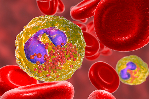
Eosinophils measure 10-16 µm in diameter, and have a characteristic bi-lobed, segmented nucleus. The presence of around 200 specific granules that contain inflammatory mediators – including major basic protein (MBP1), eosinophilic cationic protein (ECP), eosinophil-derived neurotoxin (EDN), and eosinophil peroxidase (EOP) – distinguishes them from other granulocytes.3
Function
While eosinophils can function as antigen presenting cells, their main role is to generate an inflammatory response locally and systemically against invading parasites, fungi and even viruses, through the release of cytotoxic granules. The proteins and enzymes secreted from their granules have defensive properties but, conversely, they are also linked to pathological changes and damage to healthy tissues. Apart from infections, certain myeloid hematological malignancies as well as other elevated serum levels of IL-3 and IL-5 are associated with Eosinophilia (>500 eosinophils/µl blood).4
Emerging Research
Eosinophil recruitment is well known for generating airway hyperresponsiveness in chronic respiratory diseases, making this a promising therapeutic target. For example, the drug benralizumab is directed against the IL-5 receptor α-chain, reducing the survival of eosinophils, and is now a recognized treatment for severe eosinophilic asthma.5 Eosinophils are increasingly recognized and studied for their anti-tumorigenic effects in the tumor microenvironment, potentially making them suitable as predictive biomarkers or even therapeutic candidates.6
Tools to Study Eosinophils
Flow cytometry is a key method to study the phenotype and function of eosinophils. Other ways to study these cells are microscopy, quantification of secreted cytokines by ELISA and related multiplex approaches, as well as functional assays such as migration upon exposure to chemokines.
Cell Markers
CD66b and CD49d are recognized as general markers for eosinophils as well as the cytokine receptors expressed on their surface such as IL-5Rα/CD125, CCR3/CD193, IL-3R/CD123, and IFNGR1. Neutrophils and eosinophils jointly express CD66 and CD15, but they can be distinguished by their differential expression of CD16 – eosinophils are CD16 negative.7
Overview of Specific Markers for Eosinophils
| Surface Marker | Alternative Names | Location |
|---|---|---|
| CD15 | Lewis x | Surface |
| CD125 | IL-5Rα | Surface |
| CD123 | IL-3α | Surface |
| CD294 | GPR44, CRTH2 | Surface |
| CD193 | CCR3 | Surface |
| FcεR1 | high affinity IgE receptor | Surface |
| CD170 | SiglecF | Surface |
| CD35 | CR1, C3b/C4b receptor | Surface |
| CD43 | Leukosialin, Sialophorin | Surface |
| MBPs | Major basic proteins | Secreted by cell |
| EDN | Eosinophil-derived neurotoxin | Secreted by cell |
| EPX | Eosinophil peroxidase | Secreted by cell |
| CD66b | CEACAM8, CGM6, NCA-95 | Surface |
| Siglec 8 | Surface | |
| CD49d | Integrin alpha 4 | Surface |
| CD116 | GM-CSFR alpha chain | Surface |
References
1. Metcalfe, D.D., Pawankar et al. (2016). Biomarkers of the involvement of mast cells, basophils and eosinophils in asthma and allergic diseases. World Allergy Organ J. 9:7. https://www.worldallergyorganizationjournal.org
2. Ramirez, Giuseppe A et al. (2018) “Eosinophils from Physiology to Disease: A Comprehensive Review.” BioMed Research International vol. 2018 9095275. https://www.hindawi.com
3. McBrien Claire N., Menzies-Gow Andrew. (2017) The Biology of Eosinophils and Their Role in Asthma. Frontiers in Medicine. 4:93, https://www.frontiersin.org
4. Bain BJ. (2010). Myeloid and lymphoid neoplasms with eosinophilia and abnormalities of PDGFRA, PDGFRB or FGFR1. Haematologica. 95(5):696-8. https://haematologica.org PMID: 20442440; PMCID: PMC2864371
5. Lee LY, Hew GSY et al. (2021) Targeting eosinophils in respiratory diseases: Biological axis, emerging therapeutics, and treatment modalities. Life Sci. 15(267):118973. https://www.sciencedirect.com
6. Reichman H, Itan M. et al. (2019). Activated Eosinophils Exert Antitumorigenic Activities in Colorectal Cancer. Cancer Immunol Res. 7(3):388-400. https://aacrjournals.org
7. Simon H, Yousefi S et al. (2020) The Cellular Functions of Eosinophils: Collegium Internationale Allergologicum (CIA) Update. Int Arch Allergy Immunol .181:11-23. https://www.karger.com
