Pushing the boundaries of conventional high-complexity clinical cytometry

by Dr Carsten Lange
Flow cytometry is a powerful technique for the detailed analysis of complex populations which, over the last two decades, has evolved from a staple technique of the research laboratory into an essential part of the modern clinical laboratory.
Some of the current boundaries for Clinical Flow Cytometry include its critical use for phenotyping hematological malignancies, as well as playing a vital role — along with other testing methods — in diagnosing disease, informing treatment plans, and monitoring patients.
The power to see more
The performance of flow cytometers is typically measured by their capacity to resolve and their sensitivity to detect dim and/or rare populations. In this regard, efficient light management for optimal excitation and emission of fluorochrome-tagged cells is critical to performance.
APD technology
Light is managed through the Wavelength Division Multiplexer (WDM) which uses bandpass filters to deconstruct the light and efficiently deliver it to the detectors.
The innovative detectors used to measure each parameter are avalanche photodiodes (APDs), which are highly sensitive semiconductor devices. By contrast, conventional clinical cytometers to date have (and continue to use) photomultiplier tubes (PMTs).
The major advantages of using APDs over PMTs include but are not limited to:
- Enhanced linearity;
- 4 — 5 times the quantum efficiency;
- Higher dynamic range, 106 versus 103;
- Smaller size
Figure 1 shows the WDM of the first commercially available clinical cytometer to use compact APDs which reduce the overall instrument footprint (DxFLEX, Beckman Coulter). Each WDM contains optical and detector components to selectively measure specific wavelengths. This improves light collection for higher sensitivity to detect dim populations.
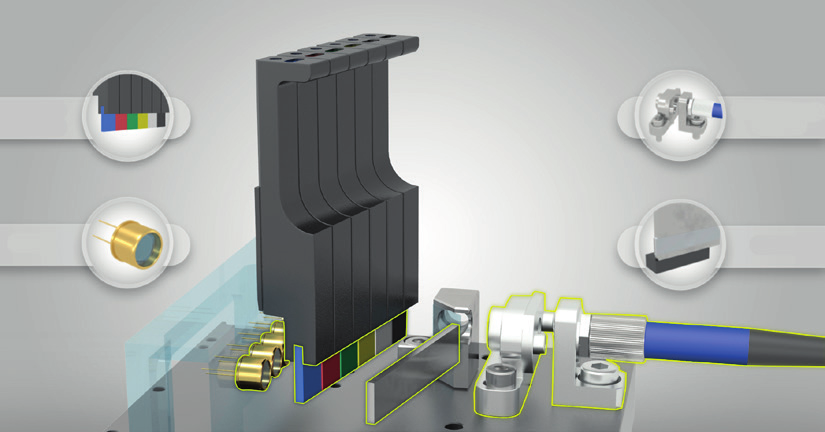
Figure 1. The WDM uses fiber optics and bandpass filters to separate the light wavelengths
The WDM’s innovative and simple design uses a single bandpass filter to select the various colors of light. This contrasts with traditional clinical cytometers, which use a series of dichroic steering filters and bandpass filters that bounce the light along an array, leading to successively less available light, resulting in diminishing light collection efficiency, and ultimately compromising fluorescence sensitivity and resolution.
Simplifying High Complexity
Leveraging the linearity of detection systems that use APDs in the operation of the cytometer can be dramatically simplified owing to the predictability of the signals. The linear gain and the normalization performed during the daily quality control routine take care of the relative variations during instrument set-up commonly seen in instruments. Further, setting up a high-complexity assay is simplified by using a software gain-only adjustment.
The linearity of gain adjustment also simplifies the typically arduous task of spectral compensation which has been the barrier for many to push to a higher number of colors/parameters. To maximize the benefit of APD linearity, new software algorithms have been developed that facilitate the setup and analysis of high-complexity experiments by simplifying compensation.

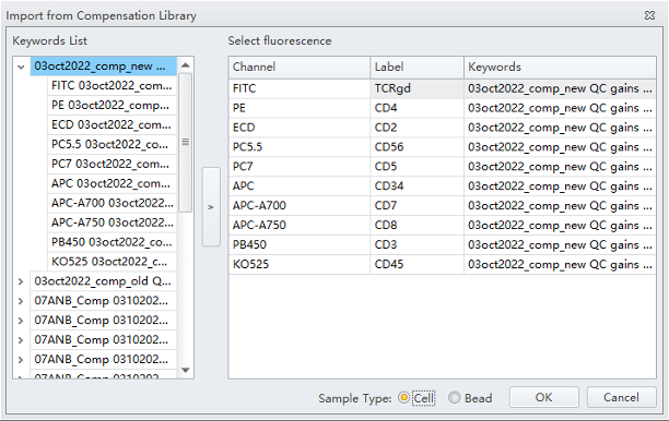
It is now possible to create a compensation library that stores the APD gain settings and spectral spill-over coefficients for every parameter and multicolor combination. This allows users to make a virtual spectral compensation matrix by selecting various single colors from the library. In addition, the library can intelligently adjust the compensation values when gains are adjusted owing to the predictive responses of linear APDs. The result is a dramatically simplified and intuitive method of setting up high-complexity applications.
The Size Factor
Saves lab space — no compressor cart. Dimensions: 28.3 in x 17.1 in x 13.4 in (72 cm x 43.5 cm x 34 cm).
The future is now
Combining powerful performance and innovative design and technology, it is possible to deliver a compact, easy-to-use flow cytometer. Pushing the boundaries of conventional flow cytometry, today’s — and tomorrow’s — cytometers simplify high-complexity applications in the clinical laboratory, as well as a deeper understanding in the frontier applications of hematopoietic cancers. Flow cytometry remains a powerful tool for interrogating complex questions. Today’s clinical laboratories want to harness that power and are demanding smaller and more powerful flow cytometers that are more affordable and easier to use. Using innovation, our engineers can deliver solutions to meet the challenge.
