Method Agreement Performance Study of the DxFLEX ClearLLab 10C Application System

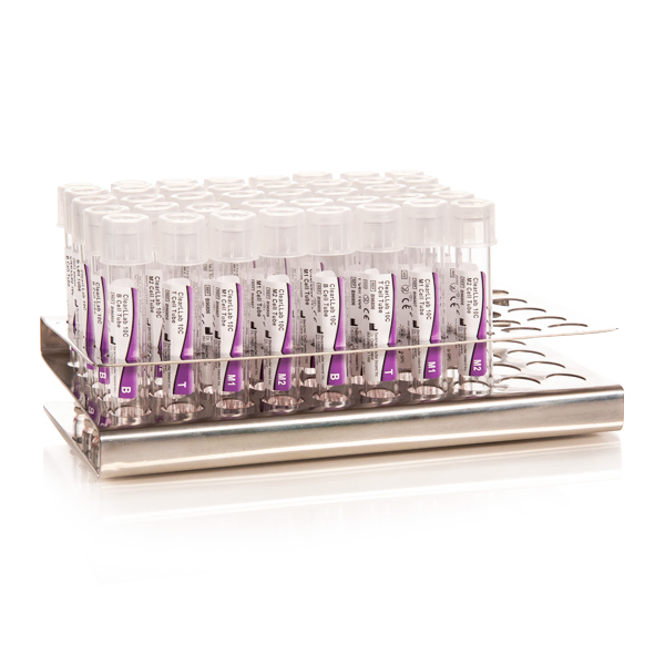
Sample Preparation
Integrated FDA cleared and CE-marked IVD leukemia and lymphoma* (L&L) immunophenotyping solution for lymphoid and myeloid lineages
*For Non-Hodgkin’s lymphoma only
Sample Acquisition
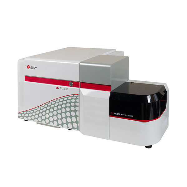
The DxFLEX Flow Cytometer streamlines high-complexity testing with a 13-color** capability and new detector technology for easier compensation.
**In the US, the DxFLEX flow cytometer is cleared for 10-color in vitro diagnostic use with the ClearLLab 10C Reagent System. Fluorescence channels FL11-FL13 and all other applications are for Research Use Only.
Sample Analysis

Flow Cytometry Analysis Software
Purpose-built for clinical laboratories, Kaluza C Flow Cytometry Analysis Software helps you turn critical data into meaningful patient reports.
Introduction
Flow cytometry immunophenotyping (FCI) is a valuable diagnostics tool for hematolymphoid malignancies.1 The DxFLEX* flow cytometer is the clinical version of the CytoFLEX instrument. Derived from the successful research platform, DxFLEX flow cytometer has the advantage of using Avalanche Photodiode (APD) technology, which has higher sensitivity than typical Photomultiplier Tube (PMT)-based cytometers and makes high-color flow cytometry testing more accessible because there is a linear correlation between gain setting / voltages and measured fluorescence intensities.2, 3
By leveraging the ClearLLab 10C** application, a multisite clinical study was conducted to demonstrate its performance equivalency between DxFLEX flow cytometers (Test Method) and Navios EX flow cytometers (Predicate Method).
* In the US, the DxFLEX flow cytometer is cleared for 10-color in vitro diagnostic use with the ClearLLab 10C Reagent System. Fluorescence channels FL11-FL13 and all other applications are for Research Use Only.
** The ClearLLab 10C Reagent System on the DxFLEX flow cytometer has received 510k clearance and IVDR certification.
Results & Discussion
A multicenter clinical trial was conducted at five (5) sites across the United States, Canada, and Europe to evaluate the clinical accuracy performance of the DxFLEX ClearLLab 10C system. A total combined 527 subjects, including 300 WB (whole blood), 172 BM (bone marrow), and 55 LN (lymph node) specimens, were enrolled. Within all WB and BM specimens, 39.62% were collected in K2EDTA, 35.59% in Heparin, and 24.79% in ACD. The distribution of the disease categories of the enrolled specimens were approximately as follow: 19% Acute Leukemias, 24% Chronic Leukemias, 36% Non-Hodgkin Lymphomas, 10% Plasma Cell Neoplasms, 8% Myelodysplasias, and 3% Myeloproliferative Neoplasms. The qualitative phenotype agreement (presence or absence of phenotypically abnormal population) from the DxFLEX 10C system (Test Method) was compared to that from the Navios EX 10C system (Predicate Method).
Statistical analysis (Table 1) indicates that for the total 527 specimens, the DxFLEX ClearLLab 10C system demonstrated 100% agreement in detecting the presence of an abnormal phenotype (246 phenotypically abnormal specimens) with lower 95% confidence limit at 98.46%, and agreed 100% in confirming the absence of abnormal phenotype (281 phenotypically normal specimens) with lower 95% confidence limit at 98.65%. This provides the overall agreement (both phenotypically normal and abnormal) at 100% with 95% confidence limit at 99.28% between both systems.
| DxFLEX (Test Method) | Navios EX (Predicate Method) | Sum | |
| Presence of Abnormal Phenotype (+) | Absence of Abnormal Phenotype (-) | ||
| Presence of Abnormal Phenotype (+) | 246 | 0 | 246 |
| Absence of Abnormal Phenotype (-) | 0 | 281 | 281 |
| Sum | 246 | 281 | 527 |
| Agreement | Estimate | 95% Confidence Limits | |
| Lower | Upper | ||
| PPA | 1.0000 | 0.9846 | 1.0000 |
| NPA | 1.0000 | 0.9865 | 1.0000 |
| OPA | 1.0000 | 0.9928 | 1.0000 |
Table 1. Clinical Accuracy Performance of All Specimens.
Conclusion
The results from this multicenter clinical study demonstrated the DxFLEX 10C system is equivalent to the Navios EX 10C system for identifying the presence or absence of abnormal populations per the intended use of the ClearLLab 10C application.
Materials
- DxFLEX Flow Cytometer with CytExpert for DxFLEX software
- DxFLEX AutoLoader
- DxFLEX Daily QC Fluorospheres
- ClearLLab Compensation Kit and Compensation Beads
- ClearLLab Control Cells (Normal and Abnormal)
- ClearLLab 10C B, T, M1 and M2 Cell Tubes
- IOTest 3 Fixative Solution and Lysing Solution
- CytoFLEX, DxFLEX, IsoFlow Sheath fluid
- PBS
- Heat inactivated FBS
- FlowClean Cleaning Agent
- Contrad 70 Reagent
- Navios EX Flow Cytometer
- Flow-Check Pro Fluorospheres
- Flow-Set Pro Fluorospheres
- De-identified residual clinical specimens per intended use of the ClearLLab 10C application
- Kaluza C Analysis software
Methods
A multisite clinical study was designed per CLSI EP09c1 and was conducted to demonstrate qualitative phenotypic (abnormal or normal) agreement from using ClearLLab 10C panels on the DxFLEX vs Navios EX flow cytometers. The study included specimens of whole blood (WB) and bone marrow (BM) collected in anticoagulant K2EDTA, Acid Citrate Dextrose (ACD) or Heparin, as well as specimens of lymph node (LN), which overall reflect the three major specimen types distributed in the leukemia and lymphoma (L&L) diseased population. All tested residual specimens were approved under WCG IRB. In brief, leftover samples from routine clinical workup were screened to confirm eligibility for clinical enrollment per the intended use of the ClearLLab 10C application. The qualified residual specimens were then prepared per reagent instructions for use (IFU), and subsequently acquired once on Navios EX and once on DxFLEX flow cytometer. All products were handled based on the IFU or associated System Guide. Kaluza C Analysis software was used for further immunophenotyping evaluation. A pathologist or equivalent professional from each site, blinded to the clinical diagnosis, assessed for the presence or absence of an abnormal phenotype per CLSI H43-A2.2 In addition, a detailed phenotype description was provided if an abnormal cell population was identified from Navios EX 10C system and/or DxFLEX 10C system.
Qualitative agreement was assessed by immunophenotyping of the presence or absence of abnormal phenotype identified with the ClearLLab 10C Panels on the DxFLEX flow cytometer (Test Method) vs Navios EX flow cytometer (Predicate Method). Positive Percent Agreement (PPA), Negative Percent Agreement (NPA) and Overall Percent Agreement (OPA) was calculated for all specimens combined. The two-sided 95% confidence intervals were calculated using the Score approach. SAS statistical software was used for data analysis in accordance with Clinical and Laboratory Standards Institute CLSI EP12-Ed3.3
References
- Craig, F. E. and K. A. Foon (2008). "Flow cytometric immunophenotyping for hematologic neoplasms." Blood 111(8): 3941-3967.
- Hedley BD, Cheng G, Keeney M, et al. A multicenter study evaluation of the ClearLLab 10C panels. Cytometry Part B: Clinical Cytometry. Published online 2020.
- Lawrence WG, Varadi G, Entine G, Podniesinski E, Wallace PK. Enhanced Red and Near Infrared Detection in Flow Cytometry Using Avalanche Photodiodes. Cytometry A. 2008;73(8):767-776. doi:10.1002/cyto.a.20595
- CLSI. Measurement Procedure Comparison and Bias Estimation Using Patient Samples; Approved Guideline – Third Edition. CLSI document EP09c. Wayne, PA: Clinical and Laboratory Standard Institute; 2018.
- CLSI. Clinical Flow Cytometric Analysis of Neoplastic Hematolymphoid Cells – Second Edition. CLSI document H43-A2. Wayne, PA: Clinical and Laboratory Standard Institute; 2007.
- CLSI. User Protocol for Evaluation of Qualitative Test Performance; Approved Guideline – Third Edition. CLSI document EP12-Ed3. Wayne, PA: Clinical and Laboratory Standard Institute; 2023.
